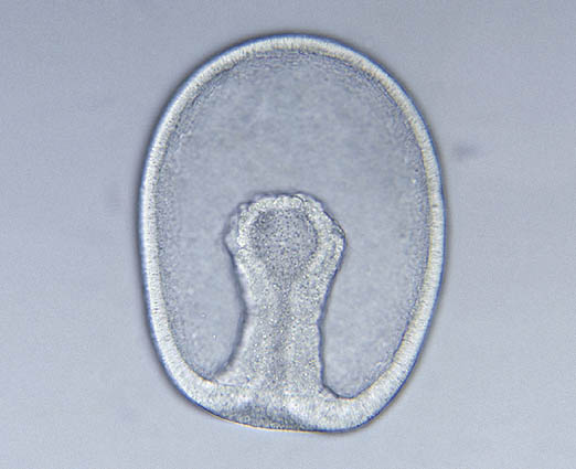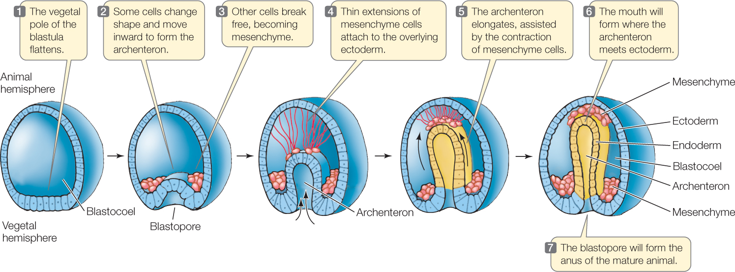

Much of this initial inbending is due to cell shape changes, and later in gastrulation convergent-extension occurs to further extend the length of the archenteron. sea urchins when this is the case, it displays incomplete adaptation. (1962) and Galileo GROWTH FACTORS AND EARLY MESODERM MORPHOGENESIS 159. Refer to figure 12 in Gastrulation in Amphibians chapter. 07 Gastrulation Biology Biological Processes Zoology. A Gamer Thing Rak Tika Aether Currents Alberto. 2 Dog Friendly Restaurants In Sea Girt Nj Bringfido Frate. As the IMZ involutes, it also extends posteriorly and converges around the circumference of the blastopore, but does so inside, out of sight.

Small micromeres are believed to contribute to the germline in the sea urchin (Juliano et al. Cells are rolling around the blastopore from outside to in, apically constricting and subducting.
#Sea urchin blastopore movie
Movie13_4 Xenopus laevis Open faced explant: (40 minutes elapsed) A timelapse confocal movie of two fluorescently labeled cells shows the characteristic mediolateral elongation and polarized protrusive activity of intercalating deep mesodermal cells. Gastrulation is a fundamental phase of animal embryogenesis during which germ layers are specified, rearranged, and shaped into a body plan with organ rudiments. Credit: David Shook, Movie 19_3 Xenopus laevis Keller Sandwich: (9 hours elapsed, 45 minutes/second): Vegetal is down, animal is up explant starts at about stage 10. Found inside – Page iiiThe purpose of the present book is to present, in a complete and orderly fashion, the enormous amount of information which has been gathered, in the course of a hun dred years of sea urchin embryology. Gastrulation in Gallus gallus (Domestic Chicken).
#Sea urchin blastopore skin
In the chick embryo, the cells of the ectoderm go on to form the skin and neural tissue, endoderm cells line the respiratory and gastrointestinal tracts, and the kidneys, circulatory system and skeleton are made from the mesoderm cells.

Later, the cells of Hensen’s node regress, paving the way for the formation of the central nervous system. The cells making up Hensen’s node at the end of the primitive streak elongate across the blastula. One of the unique features of chick gastrulation is the cellular rearrangement that occurs at the posterior end of the blastula and forms the primitive streak, a thickening of the tissue. The lip is the point where the cells begin to turn and migrate inward, forming the blastopore. Another interesting aspect of frog gastrulation is that the blastopore forms a “lip” exactly 180 degrees opposite from where the sperm entered the egg. Also different, is that the cells of the blastula in the frog form the ectoderm or endoderm while the mesoderm is made from the yolk cells inside.

One of the main differences is that the blastula is not hollow but is filled with yolk cells. Gastrulation in the frog is similar to the sea urchin, but it’s more complicated. In sea urchins, all three primary germ layers originate from the same outside layer of cells. The anus forms at the spot where the invagination started on the surface. The archenteron elongates and eventually fuses with the epithelial cells on the surface to form the mouth. Then, the archenteron (primitive gut) forms as the blastocyst invaginates creating the blastopore. In the first step of gastrulation, primary mesenchyme cells use chemical cues inside the blastula to migrate to the inside of the sphere where they will eventually fuse and form the larval skeleton made of spicules formed from calcium carbonate. Gastrulation in sea urchins is used as a starting point for understanding the process, as it is less complicated or more “simple” compared to other species, and the process only takes about 9 hours. Frogs, chickens, and sea urchins are 3 species most studied by developmental biologists and comparative embryologists.įigure 1: The image above shows the process of transformation from a single-celled zygote to a gastrula.įigure 2: The image above shows how gastrulation changes the number of cell layers from one to three. Each species has its own uniqueness when it comes to the process of gastrulation, but there are similarities that span the entire animal kingdom. The layers are called the primary germ layers the endoderm, ectoderm, and mesoderm (Figure 2). This is a critical point in development because it is when the embryo transforms itself from a hollow sphere made from a single layer of cells into a multi-layered structure. It does this by folding itself inward as shown in Figure 1. Gastrulation is a phase in the embryonic development of animals where the blastula reorganizes itself into a gastrula.


 0 kommentar(er)
